PLANKTON
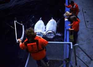
Plankton Tow. Courtesy of Scripps Institution of Oceanography
Paired plankton nets are cast over the side to collect plankton, principally zooplankton such as copepods, arrow worms, and invertebrate larvae. The pair of nets (known as “Bongo nets”) provides some degree of replication for spatial sampling. Note the sampler at lower left, who is holding a protracter that enables her to estimate the angle of entry of the wire holding the nets; with this and a measure of the line that is paid out, one can get an estimate of the sampling depth. Most plankton nets are equipped with propeller- driven anemometers, in order to estimate the linear distance towed. With the cross-sectional area, this allows the volume of water sampled to be estimated. Phytoplankton can be sampled with nets, but they are usually taken with pumps or automatically closing bottles.
These samples were taken from the R.V. New Horizon, which sails out of the Scripps Institution of Oceanography located in La Jolla, California.

R. V. New Horizon. Courtesy of Scripps Institution of Oceanography
This vessel was used for sampling in the previous slide. It is equipped for a wide variety of oceanographic sampling methods.
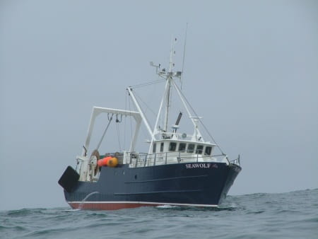
R/V Seawolf, Stony Brook University
This is a smaller vessel, used by the School of Marine and Atmospheric Sciences at Stony Brook University, for sampling in estuarine and continental shelf waters. Note the rear “Inverted-U” frame boom for deploying sampling equipment.

Satellite Image of Gulf of Maine: Chlorophyll. Data collected by E. Feldman
Satellites can be equipped with color scanners that can efficiently detect variation over very large areas. This image was produced from data collected by the Coastal Zone Color Scanner over the Gulf of Maine. The color red is a false color, indicating high concentrations of chlorophyll. Note the strong concentration over Georges Bank (GB), one of the most productive fishing grounds in the world (at least until overfishing occurred in recent decades).
While the color gives us very useful ideas of changes in chlorophyll concentrations, it must be calibrated by measurements made “on the ground.” Many problems still prevent a universal calibration, including the problem of detecting chlorophyll at depth and the relationship of total chlorophyll, a measure of biomass, to productivity.


Ditylum brightwellii (left), Thalassiosira sp. Photos by George Rowland
Common diatoms in the spring phytoplankton bloom of temperate- boreal waters. Diatoms have an external skeleton of silica and silicon may therefore be a limiting element to diatom population growth. This species occurs in chains of a few cells.
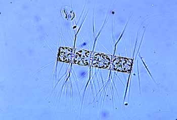
Bacteriastrum hyalinum. Photo by George Rowland
This diatom occurs in chains of cells, armed with spines. This diatom is not preferred by suspension feeding bivalve mollusks. Have you any ideas why the spines are present?

Asterionellopsis glacialis. Photo by George Rowland
This diatom is common in the phytoplankton and often a dominant form.
Ceratium. Photo by George Rowland
Dinoflagellates are important parts of the phytoplankton in estuarine and shelf waters. They reproduce by oblique cell division and often come to dominate the summer phytoplankton in temperate waters. Like many other dinoflagellates, Ceratium can produce cysts that may rest dormant within the sediment. This one is about 250 micrometers long.

Protoperidinium. Photo by George Rowland
This is a dinoflagellate common in estuarine and shelf waters. It is about 50 micrometers across. It is heterotrophic, not deriving sustenance by means of photosynthesis.
Pfiesteria piscicida. Courtesy of JoAnn Burkholder
It seems extreme to name a dinoflagellate species piscicida, or fish killer, but that is exactly the effect that this newly discovered dinoflagellate has on estuarine fishes. P. piscicida produces a toxin that kills fishes. This organism is heterotrophic and, once the toxin has attacked the surface of the fish, it feeds on the disaggregated and decomposing fish carcass. Moreover, it appears that the dinoflagellate responds to the presence of fish, especially in sluggish estuarine waters rich in phosphate. Cyst stages in the sediment are activated to a stage that attacks the fish.
Research by JoAnn Burkholder suggests that this dinoflagellate species has several stages, with distinct morphologies. Some resemble the typical biflagellate dinoflagellate form, whereas others are amoeboid in form.
This species has been implicated in sudden fish kills in North Carolina estuarine waters in recent years, particularly in Pamlico Sound, which is the second largest estuary on the east coast of the United States. Toxic algal blooms are on the rise throughout the world in recent years.
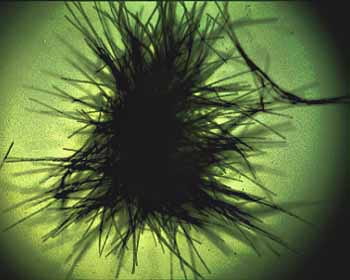
Trichodesmium thiebautii. Photo by Edward Carpenter
The radial form of the filamentous blue green that is common in tropical and subtropical oceans. Like other cyanobacteria, this species is a nitrogen fixer. It occasionally occurs in dense mats on the sea surface, which may color the sea water. This organism is believed to be the source of the name “Red Sea.”


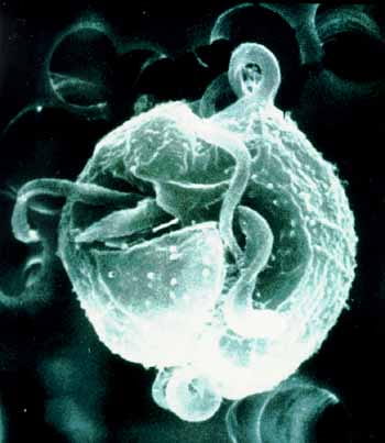
Ciliate Strombidium capitatum. Photos by Diane Stoecker
This ciliate, about 30 micrometers in size, is common in shelf and estuarine environments and is one of the protistans important in the microbial loop. On the right is a light micrograph, showing oral cilia on the upper right. This organism consumes microalgae and can retain chloroplasts, which photosynthesize and release nutrients to the ciliate. The picture at left was taken using an blue end of the spectrum light source. The red dots are caused by fluorescing chlorophyll. Thus, Strombidium is both a consumer and a primary producer, or member of the mixoplankton.
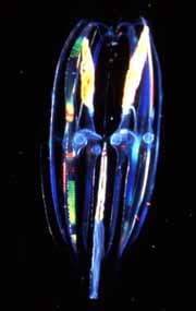
Ciliate Strombidium conicum. Photo by Diane Stoecker
This is a scanning electron micrograph of a common oligotrich ciliate that feeds on algae and retains chloroplasts and also feed on smaller protists by means of the ciliary tufts on top. These tufts operate at low Reynolds number and sweep particles into the mouth. This species does not keep endosymbiotic chloroplasts. Scale is in microns.

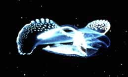
Aequorea victoria. Photo by Claudia Mills
The jellyfish Aequorea victoria is common in the Puget Sound region. It is the source of bioluminescent “green fluorescent protein”, so important in widespread molecular-cellular studies.

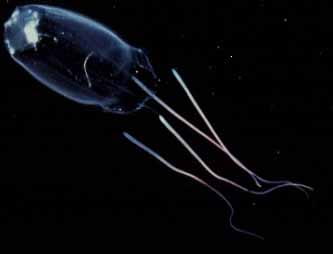
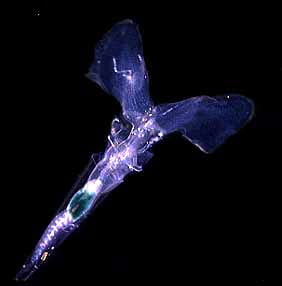
Pandea conica. Photo by Laurence P. Madin
The anthomedusan Pandea conica common in continental slope waters of the western Atlantic, and a predator on other gelatinous zooplankton. Up to 30 mm high.
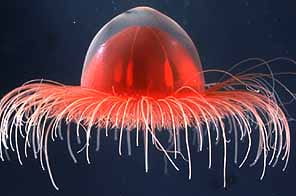
Benthocodon pedunculata. Photo by Claudia Mills
The trachymedusan jellyfish Benthocodon pedunculata was collected from a submarine at a depth of 900 m, offshore of the Bahamas.
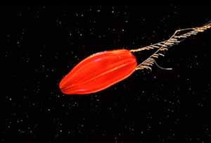
Cubozoan, Carybdea alata. Photo by Marsh Youngbluth
This jellyfish lives in subtropical waters in the upper 100 m of the water column.

Porpita
The hydrozoan Porpita has a flattened float, with tentacles that extend radially and probably serve to stabilize the animal in the water. This specimen was collected in coastal waters of North Carolina and is about 10 cm across.

Physophora hydrostatica. Photo by Laurence P. Madin
Physophora is a siphonophore, common in the tropical ocean. Siphonophores are holoplanktonic and include the Portuguese Man of War. Physophora feeds upon phytoplankton and smaller zooplankton, such as herbivorous copepods. Colonies are up to 50 mm high.

Bolinopsis vitrea. Photo by Laurence P. Madin
Bolinopsis vitrea, a lobate ctenophore, common in the Caribbean and other subtropical regions, is ca. 60 mm high. It preys on copepods and other small crustaceans. Ctenophores lack the nematocysts characteristic of jellyfish.
Unidentified Ctenophore. Photo by Marsh Youngbluth
This undescribed ctenophore has remarkable coloration. Ctenophores are commonly multi-colored, owing to refraction along meridional rows of fused cilia known as comb rows.
Ctenophore. Photo by Marsh Youngbluth
Ctenophores are ubiquitous in the ocean and their systematics are poorly understood. They are efficient suspension feeders, feeding on smaller zooplankton, fish eggs, and planktonic larvae.
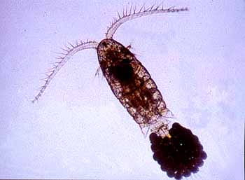
Copepod Eurytemora affinis. Photo by Carol Eunmi Lee
The herbivorous copepod Eurytemora affinis is common in estuarine waters all over the northern hemisphere. This is a female, about 1 mm long.
Populations occur in a wide range of salinities. For example, populations can be found, apparently isolated from each other, in many estuaries in Europe and in North America. Carol E. Lee has been investigating the problem of whether these low salinity populations are one genetically similar group, adapted to low salinity that have dispersed all over the world, or whether the low-salinity populations arose many times independently, perhaps evolving from fully marine ancestors. DNA sequencing evidence thus far supports the independent origin hypothesis.
Copepod Pseudodioptomus inopinus. Photo by Carol Eunmi Lee
This copepod is a detritivore and also feeds upon phytoplankton. The species is common in Korean, Chinese, and Japanese coastal waters. This female is about 1.5 mm long.
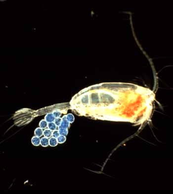
Paraeuchaeta elongata. Photo by Steve Bollens
An egg-bearing female of the copepod Euchaeta elongata, which was collected in Dabob Bay, Washington. It is carnivorous, feeding on smaller species of copepods.
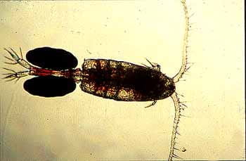
Paraeuchaeta norvegica. Photo by Jeannette Yen
This copepod is a carnivore, feeding principally upon smaller copepod species. Note the prominent mechanosensory hairs on the large first antennae, which detect the wake of approaching and escaping prey. The wake of prey bend the mechanosensory hairs and this stimulus, if of the right magnitude, is followed by an attack. When the prey are detected the copepod moves rapidly forward and can seize the prey with its maxillary appendages, which can be seen clearly here, projecting laterally from each side.
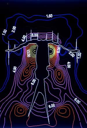
Euchaeta rimana flow field. Photo by David Fields
Although there may be turbulence around the copepod, the flow near the body surface is more regular, as this diagram of flow field intensity shows. Near the copepod the flow is laminar and viscosity is a dominant feature of flow. Thus, movements such as those of prey are dissipated near the animal and are detected by mechanosensory hairs.
The flow field was determined by placing three video cameras at right angles to each other, and measuring the velocity of particles around the copepod. The flow field is created primarily by beating of the second antennae.
For further information see paper by D. Fields and J. Yen, Bulletin of Marine Science, v. 53, pp. 84-95 (1993).
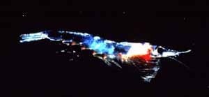
Meganyctiphanes norvegica. Photo by Marsh Youngbluth
Krill are planktonic crustacea, members of the Euphausiacea that tend to occur only in oceanic environments. They are large for planktonic crustacea, often reaching lengths of 15-20 mm (Meganyctiphanes is ca. 30 mm long). They often dominate the zooplankton in the eastern Pacific and in the Antarctic, where they are famous as principal prey for baleen whales.

Themisto compressa. Photo by Marsh Youngbluth
While most amphipods are benthic, the Hyperidean amphipods are exclusively planktonic, though they rarely are a major component of the zooplankton. They are often associated with, and feed upon, gelatinous zooplankton.

Stalked barnacle, Lepas. Photo by Rudolph Scheltema
Stalked barnacles of the genus Lepas are pelagic and are attached to floating objects, such as pieces of wood or floating seaweed.

Portunus sayi, the Sargasso Crab. Photo by Rudolph Scheltema
This crab is associated with Sargassum weed, which floats in the open western tropical Atlantic and harbors a large community of associated animals.
Gleba cordata. Photo by Marsh Youngbluth
These shelled pteropods are holoplanktonic gastropods with elegant swimming organs, or wings. Gleba cordata feeds upon phytoplankton and zooplankton that are trapped in a large free-floating mucus net.

Diacria sp. Photo by Rudolph Scheltema
Pteropods are holoplanktonic, meaning they spend their entire life cycle in the plankton. To move about and maintain position in the water column they employ elegant swimming organs. Diacria is a shell-bearing form that feeds on particles using a mucus feeding web.
Creiseis sp. Photo by Rudolph Scheltema
Another pteropod found in plankton of the open sea.
Oxygiris sp. Photo by Rudolph Scheltema
This holoplanktonic gastropod is a heteropod and is carnivorous and hooks on the radula can seize prey such as ctenophores. This group is distinct from the pteropods and it is probably derived from a prosobranchiate ancestor. The foot has been modified into a fin-like structure that flaps to move the snail. Millimeter scale along top of photo.

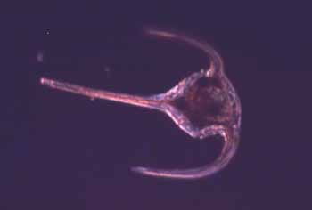
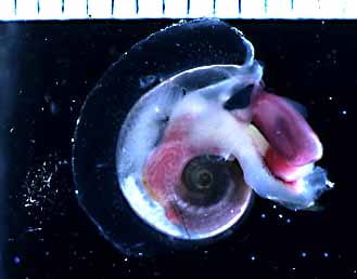
Nautilus. Photo by Peter Ward
It is not easy to classify Nautilus as plankton entirely, as it can swim in short bursts. It uses gas exchange between the chambers of its shell and the outside water to regulate buoyancy.
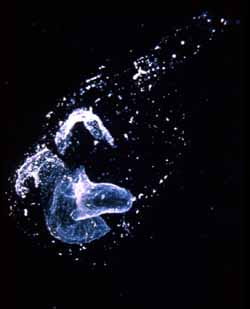
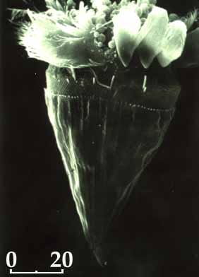
Marine Snow. Photo by Alice Alldredge
Marine snow refers to particulate organic matter that originates in the ocean. It may be formed by collisions and merging of large macromolecules and also may start from decaying material, such as the “house” of a larvacean (such as at left) or a salp colony. Marine snow often has large numbers of attached microorganisms and protistans.